Injection of Right Hip
$395.00Sagittal view of the right hip showing injection into the right hip joint.
Showing 17–32 of 61 results
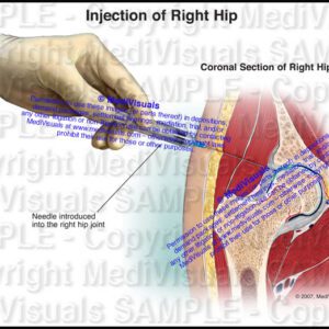
Sagittal view of the right hip showing injection into the right hip joint.
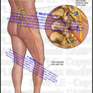
Demonstrates a disc compressing an exiting right nerve root resulting in radicular pain that radiates to the left leg.

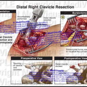
Intraoperative view of right shoulder showing an open distal clavicle resection|acromioplasty|and bursectomy.
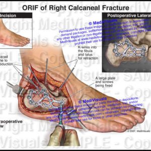
Lateral intraoperative views of right foot showing open reduction and internal fixation of calcaneus with a plate and multiple screws.
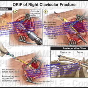
Intraoperative view demonstrating open reduction of a left clavicular fracture and fixation with a pin.
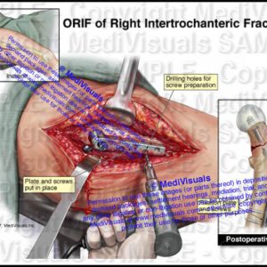
Intraoperative view of right hip with femoral neck fracture and fixation using a plate and screws.
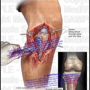
Illustrations demonstrating the surgical exposure required to reduce and fixate a tibial plateau fracture and the postoperative appearance with fixation with a plate and multiple screws.
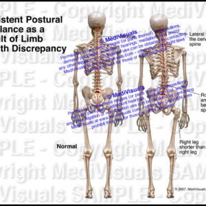
Posterior views of skeletons comparing normal stature and gait with the pelvis|spine|and head in straight alignment compared to abnormal alignment required to adjust to discrepancies in leg lengths. The abnormal alignment result in chronic spinal pain.
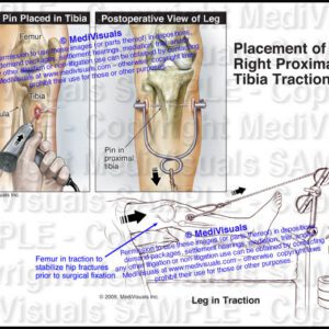
Illustrations of right leg showing the placement of tibia traction pin to realign (reduce( or relocate hip or femur fractures and dislocations.
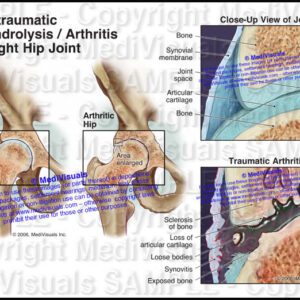
Illustrations of the right hip comparing the normal joint (with thick|shock-absorbing articular cartilage) to the joint after the development of post-traumatic degenerative changes.
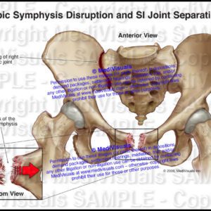
Shows an anterior view of torn pubic symphysis and disruption of the sacroiliac (SI) joint
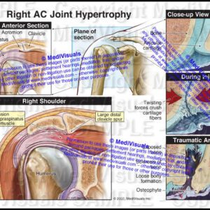
Series of illustrations comparing the normal AC joint (with thick|shock absorbing articular cartilage covering smooth articular bone) to the joint during a traumatic event and after resulting post-traumatic changes. The post-traumatic hypertrophy then results in irritation and compression of the underlying rotator cuff structures.
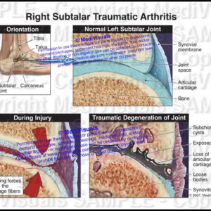
Sectional view through injured subtalar joint comparing the normal joint (with thick|shock-absorbing articular cartilage) to joint during trauma|and after the development of post-traumatic degenerative changes.