Powerful Brain Injury Exhibits
MediVisuals has developed many demonstrative evidence tools which accurately depict brain injury and corrective surgeries to effectively teach the difficult pathology involved in traumatic brain injury (TBI) cases. These include; Brain Injury Animations, Brain Injury Illustrations, Brain Models, as well as Brain Interactive Presentations and Brain 3D Reconstructions.
If you cannot find what you are looking for, or would like to use our exhibits for CLE presentations please contact us.
Brain Injury: Animations
In collaboration with some of the nation’s leading medical experts and trial attorneys who specialize in traumatic brain injury (TBI), MediVisuals has developed animations that demonstrates how traumatic brain injuries can occur, surgical procedures and mechanism of injury. Medical animations are utilized for a jury to fully understand the extent to which a plaintiff has been forced to undergo.
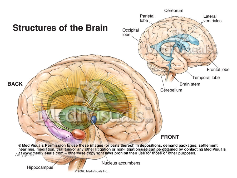
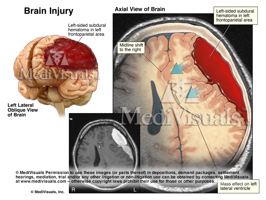
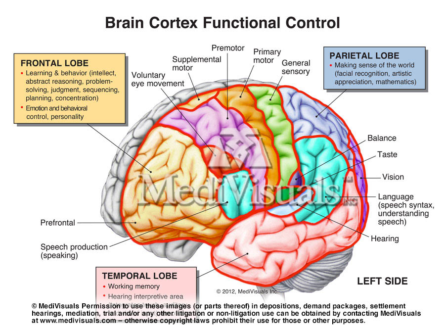
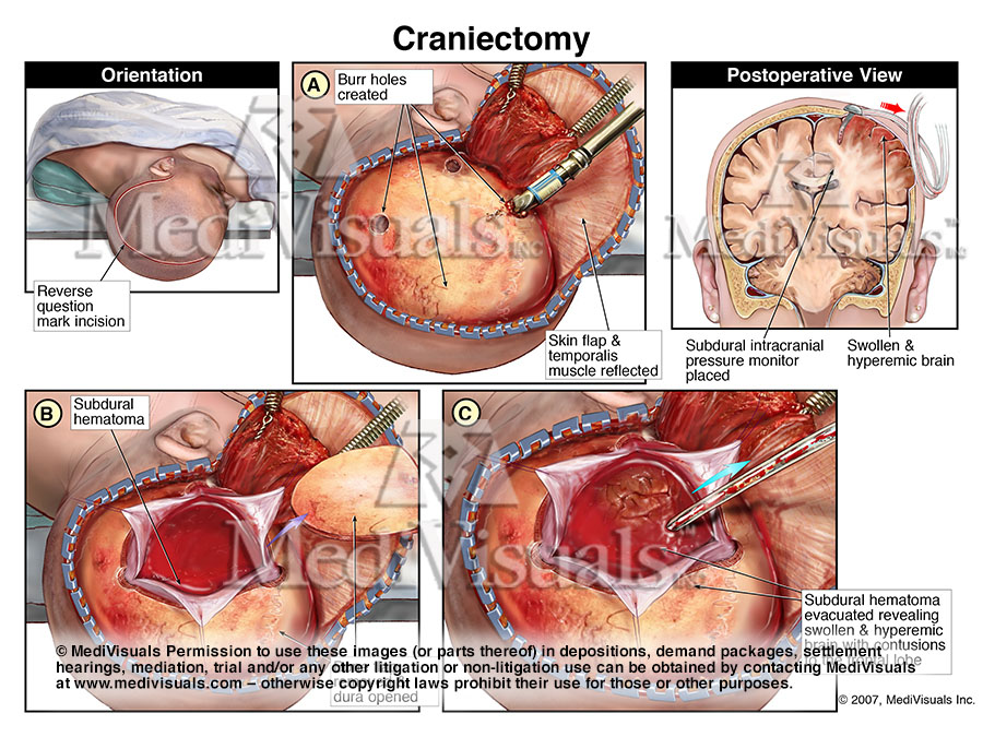
Brain Injury: Illustrations
Both stock and custom medical legal illustrations are developed by MediVisuals’ medical illustrators. Static exhibits include; medical illustrations, charts, graphs and timelines. Their experienced team of medical illustrators can modify stock exhibits to fit your needs or provide you with a complimentary consultation to develop illustrations customized to your case.
Brain Digital Interactive Presentations
Capture your audience with the most captivating, visually exciting, appealing and understandable presentations. The interactive medical legal presentations are custom-built with Powerful, Engaging, Innovative and Efficient methods that allow flexibility while delivering your message and enhancing viewer attention and comprehension. Contact MediVisuals to get started on a project.
Brain 3D Reconstruction Videos
MediVisuals’ exclusive innovative 3D diagnostic imaging technology uses CT or MRI data to show clients’ actual anatomy more clearly and accurately than ever before. The end-product may be a video or an interactive digital model which can be rotated and enlarged by the user. This FDA cleared technology is currently in use at large medical facilities.
Neuron Suicide and Murder Animation:
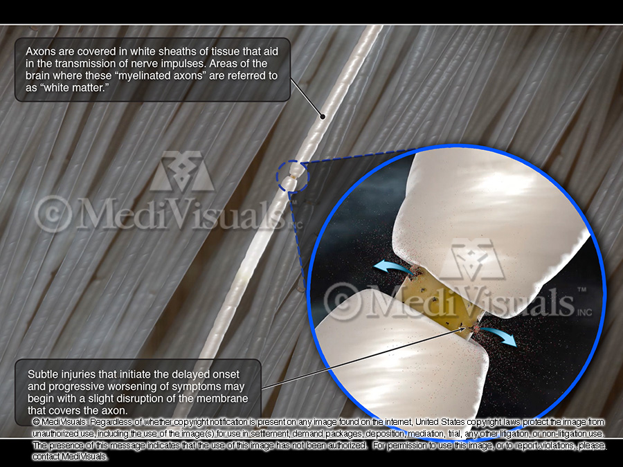
White Matter Damage
$2,495 (first case per firm)
$600 (each subsequent case per firm)
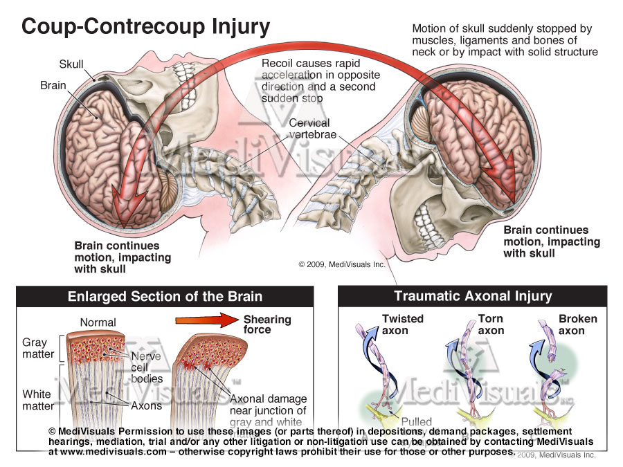
This eight-minute animation accurately and effectively explains the complexities of traumatic axonal injury and neuron “suicide and murder.” It demonstrates how incidents of TBI can initially go undetected because symptoms may not be immediately evident, and areas of injury and cell death can be too small for traditional imaging studies to detect. These symptoms may progressively worsen as the cascade of cell death continues, ultimately resulting in devastating brain injuries that do not fully develop until weeks or months after the initial incident.
Brain Injury Animations: Options for TBI Cases with LITTLE or NO Evidence of Injury on CT or MRI:
Diffuse Axonal Injuries: Invisible on MRI but Present on DTI, FA, or Tractography
Demonstrates the true appearance of abnormal findings on FA, DTI, or Tractography imaging.
The Ongoing “Process” of a TBI
Explains how TBIs are “not an event but an ongoing process” resulting in continuing axonal injuries and symptoms for months, years, or even the plaintiff’s lifetime. Full narration (with citations) as an eductional tool can be turned off for litigation.
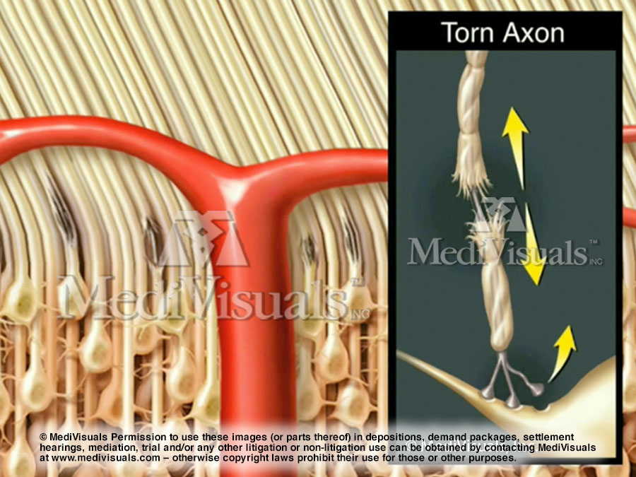
Axon Vulnerability
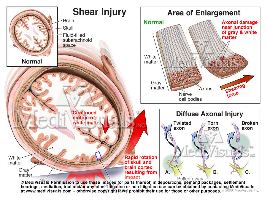
Simply and understandably emphasizes the delicate nature of axons and how vulnerable they are to shearing.
Microscopic Axonal Injuries Associated with Abnormal DTI, FA, and Tractography Findings
Demonstrates normal waterflow in and through the axons to the abnormal flow of water in areas of axonal injury that result in abnormal findings in DTI, FA, Tractography, etc.
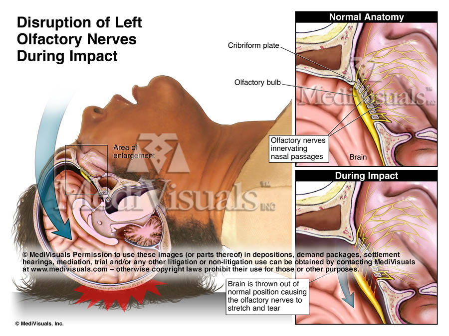
“Mild” Traumatic Brain Injury
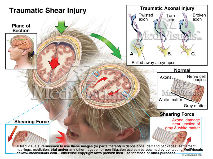
Helpful in showing and explaining shear injury from a frontal impact and how brain injuries can occur without evidence of a blow to the head. Full narration for can be turned off for litigation.
“Mild” Traumatic Brain Injury
Helpful in showing and explaining shear injury from a rear impact and how brain injuries can occur without evidence of a blow to the head
Animation Stills: Click On Image to Enlarge
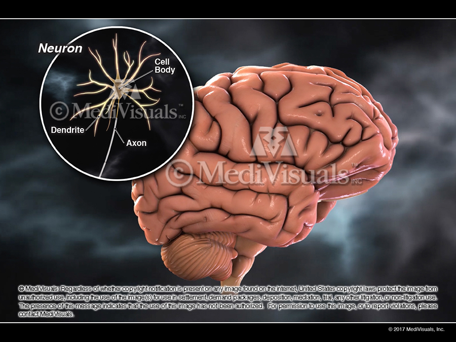
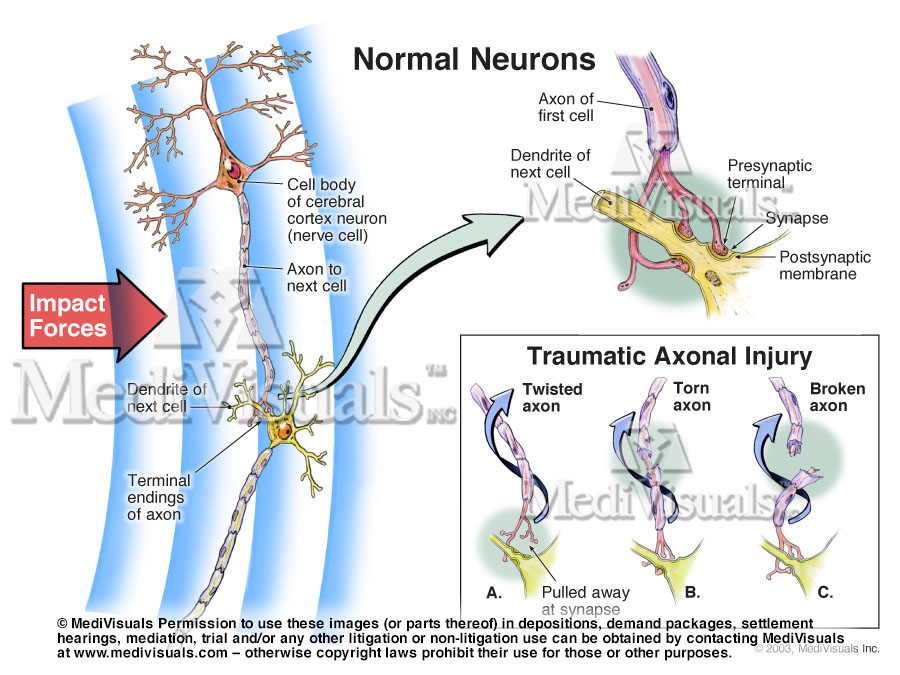
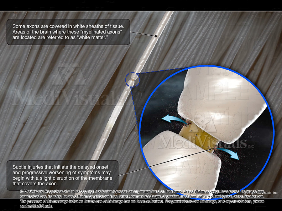
Close up of a neurons anatomy
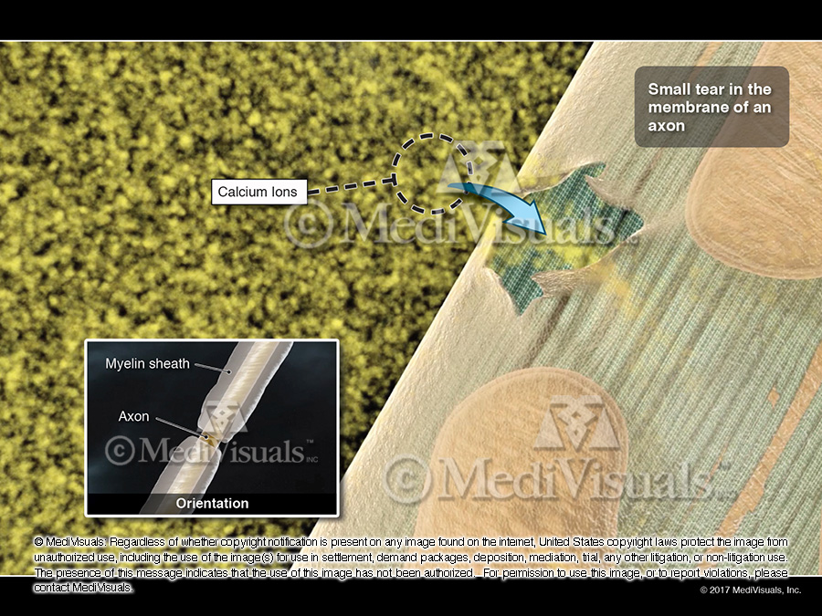
Calcium ions flowing uncontrollably into a small tear in the membrane of an axon.
Close up of an injured axon and the release of self-destructive substances.
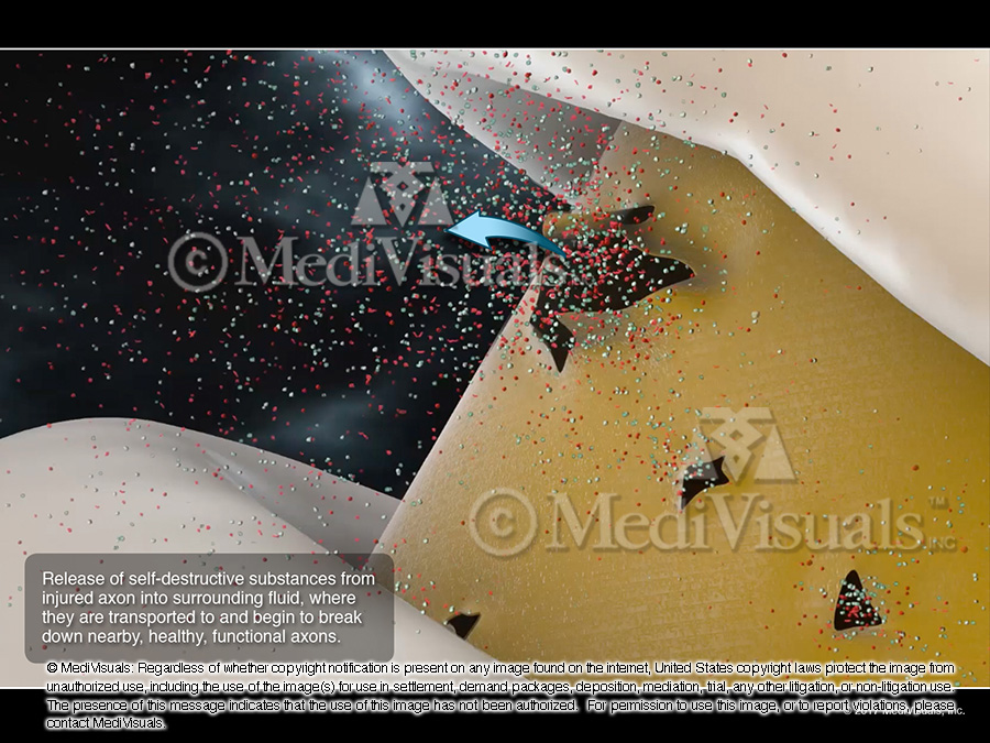
Injured axons release self-destructive substances, they begin to break down nearby, healthy, functional axons.
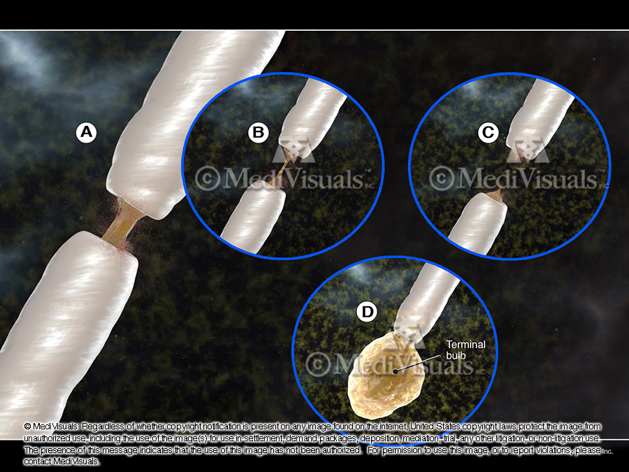
Destructive electrical and chemical impulses being discharged from a terminal bulb.

Multiple microscopic islands of injured axons, each consisting of axons – all too small to be visible on typical MRIs.
Animations are useful in effectively illustrating and assisting the jury in understanding the technical and complex issues often present in brain injury cases. It is highly likely the defense will object to getting the demonstrative evidence admitted for being more prejudicial than probative. Medivisuals has created still exhibits that can be utilized, should issues arise with getting the animation admitted into evidence. They can be purchased individually at the archive prive of $395* each or if bought in conjunction with the Neuron Suicide & Murder Animation the price is $100* each.
* price is for a digital 150 dpi jpeg file. Additional fees may occur for printing, mounting, laminating and shipping.
Brain Injury: Digital Interactive Presentations
Stock or Customized Versions
ACTUAL Anatomy. ACTUAL Evidence.
MediVisuals’ exclusive NEW! innovative 3D diagnostic imaging technology uses CT or MRI data to show clients’ actual anatomy more clearly and accurately than ever before. The end-product may be a video or an interactive digital model which can be rotated and enlarged by the user. Both formats are easily understood by laypeople. This FDA cleared technology is currently in use at large medical facilities.
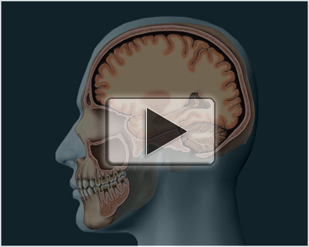
Life-Size Soft Brain Model | Click to play narrated demonstration
Price: $1295.00
MediVisuals’ soft brain model is perfect for demonstrating the weight, flexibility, and vulnerability of the human brain. It is very effective in explaining how a brain injury can occur even without evidence of a significant blow to the head. Life-size and made of silicone, the brain is soft to the touch and fits inside a standard skull. Clear plastic skull and carrying case are included with your purchase.
MediVisuals’ soft brain model is also a great educational tool for clinicians, professors, and grade school teachers.